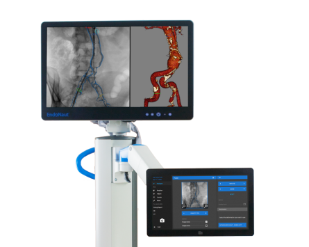
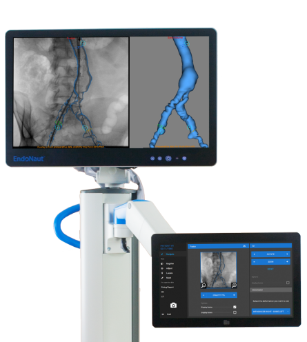
EndoNaut Aorto-Iliac
3D Roadmaps in any Operating RoomEndoNaut brings the image fusion technology into your operating room.
Have the most from the preoperative patient CT during the procedures at your fingertips. Easily and quickly combine with any mobile or fixed C-arm for a fraction of the cost of a hybrid room.
IMAGE FUSION MADE PHYSICIAN, PATIENT, I.T., OR TEAM, BUDGET-FRIENDLY
Download the brochureReliable 3D Roadmap without additional contrast.
Designed to upgrade your Operating Room
Better quality care at a lower cost.
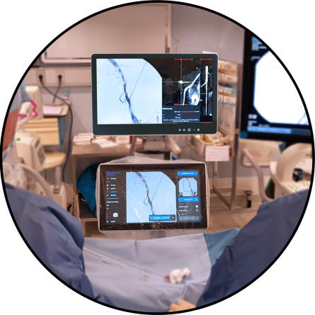
Bring image fusion technology into any existing environment. EndoNaut is already compatible with your mobile or fixed C-arms, and can be used across multiple operating rooms.
No I.T. constraints
Just connect the EndoNaut to the C-arm video output.
It's immediately ready for use.
Time-efficient
Streamlined workflow
Optimize your workflow by utilizing the full potential of your EndoSize-based 3D case planning.
Keep control
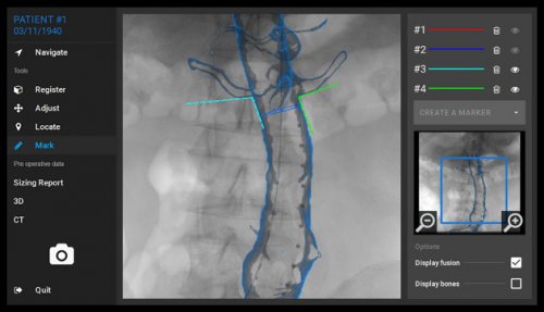
The intuitive touchscreen interface is controlled by the physician right from the operating table. Get the information you want when you need it by yourself.
Ease of use
EndoNaut has been designed to bring user-friendly 3D navigation to your fingertips. Keeping the focus on patient care.
Patient and medical staff friendly
Decreased X-ray exposure
3D overlay allows to lower procedure time and reduce contrast angiography, which contributes to a decrease of X-ray exposure for both patients and staff.
Minimum contrast agent injections
Use of 3D fusion imaging is associated with a significant reduction of nephrotoxic iodinated contrast use, limiting risks for patients with renal failure. [1]
[1] S. R. Goudeketting et al., “Pros and Cons of 3D Image Fusion in Endovascular Aortic Repair: A Systematic Review and Meta-analysis,” J. Endovasc. Ther. Off. J. Int. Soc. Endovasc. Spec., p. 1526602817708196, May 2017.
Want to see EndoNaut in real conditions?
Attend a workshop!Empowering features
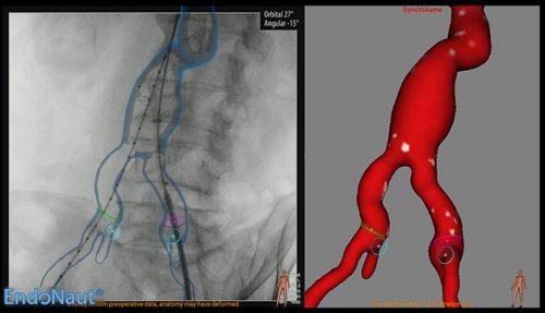
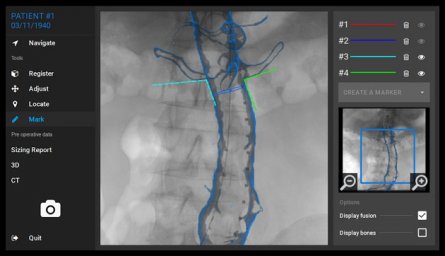
3D localization, in real time
Locate your tools in relation to the native CT scan and 3D images
Measurement
Perform length and diameter measurements on CT scan right from the operating table
Stiff wire simulation
Use pre-computed stiff wire simulation to predict and overlay the deformed vessels
2D digital zoom
Zoom on fluoroscopy without changing C-arm field-of-view to save X-ray doses
Automatic adjustment
Automatically adjust the 3D overlay based on contrast angiography
Sizing report
Access all preoperative measurements, device selection, comments, and snapshots
Custom markers
Draw markers directly on the fluoroscopy and never lose important locations
C-arm gantry planning
Simulate virtual gantry angulations to avoid parallax effects